
What Can a Keyence Microscope Reveal About Weld Defects?


Weld defects are not always obvious during routine inspection. A Keyence Microscope can help specialists examine small surface features, document damage, and identify details that warrant further testing. At Welding & Joining Management Group (WJMG), digital microscopy supports our broader work in welding evaluation, materials testing, and failure investigations.
What Is a Keyence Digital Microscope?
A Keyence digital microscope is an optical imaging system that magnifies, captures, and measures features on material surfaces. WJMG uses the Keyence VHX-5000, with magnification ranging from 20X to 1,000X, to examine specimens in two and three dimensions.
Digital microscopy provides detailed images and measurements that help document a specimen’s condition. Specialists interpret these findings alongside the material’s history and laboratory testing results.
What Weld Defects Can Digital Microscopy Help Identify?
A Keyence Microscope can help identify surface irregularities and examine material characteristics that may indicate welding problems. Visible features depend on specimen preparation and condition.
Common examination areas include:
- Surface cracks: Magnification can reveal crack openings, branching, and surrounding surface changes.
- Porosity: A prepared weld cross-section may expose pores and help document their size and distribution.
- Corrosion and pitting: Surface imaging can show localized metal loss and irregular damage patterns.
- Fracture surfaces: Magnified views can help document features associated with a broken component.
- Weld microstructure: Prepared and etched samples can reveal characteristics of the weld metal, fusion boundary, and heat-affected zone.
These observations can help characterize potential defects, but microscopic images alone cannot establish why a problem developed.
How Does 3D Imaging Support Weld Examination?
Uneven weld edges, pits, and fractures can be difficult to evaluate in two dimensions.
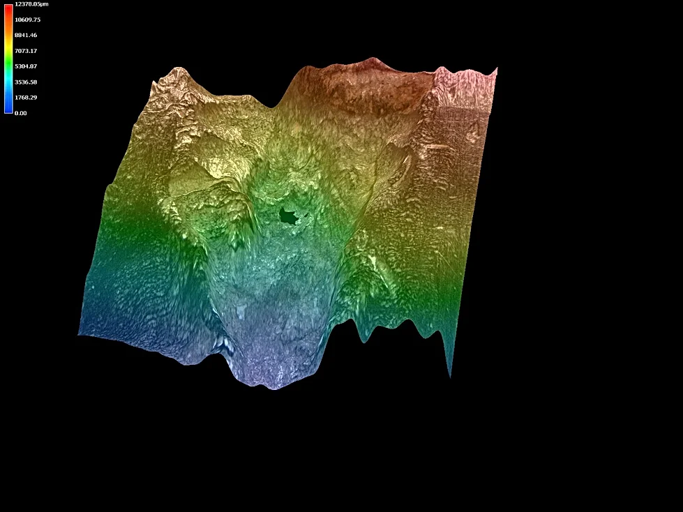
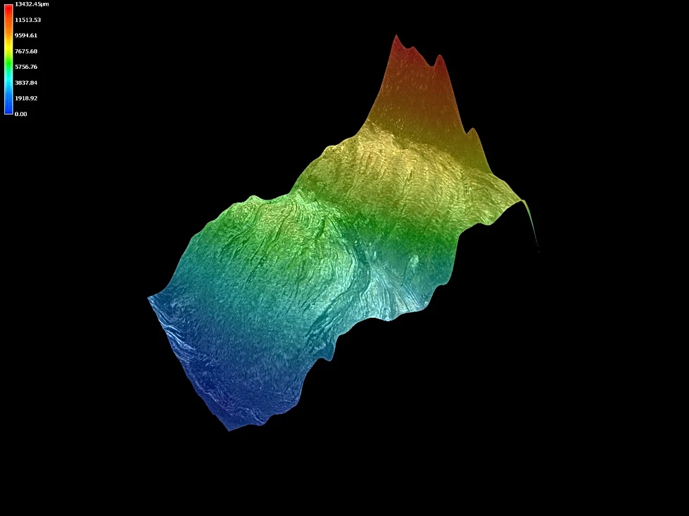
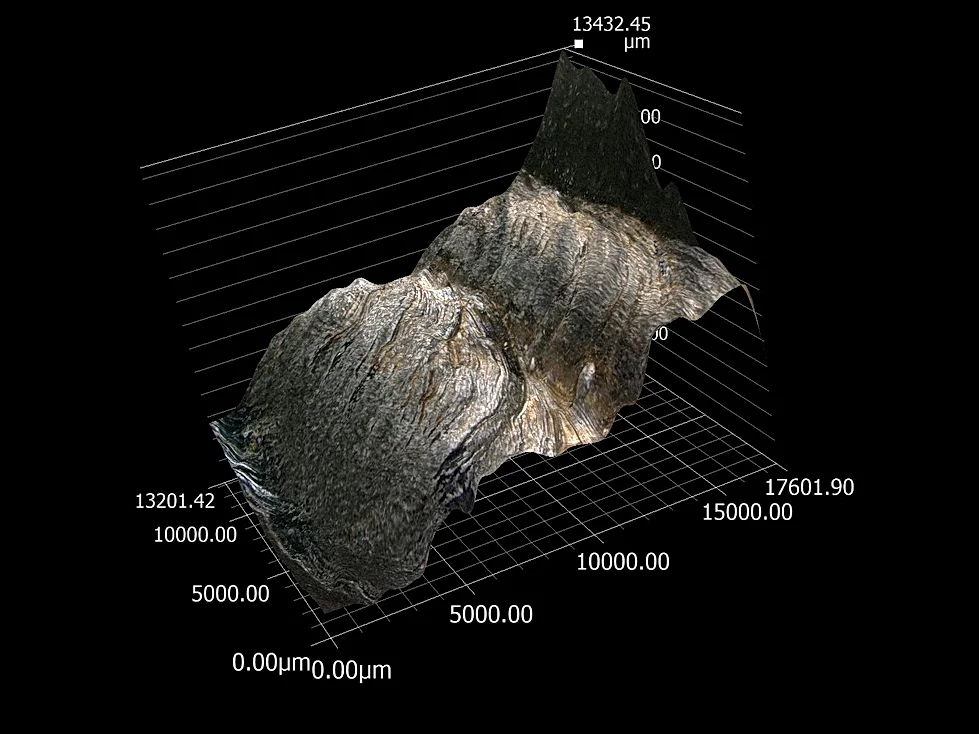
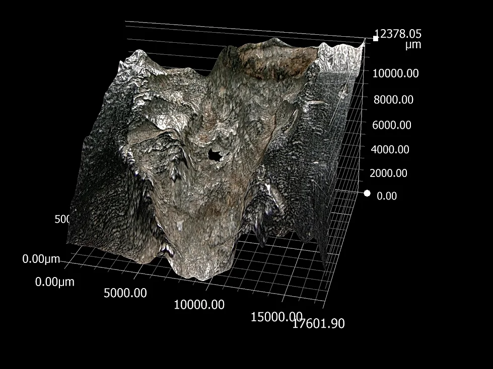
A Keyence digital microscope uses three-dimensional imaging to capture surface height information, helping specialists visualize profiles and measure accessible features. Depth-composition techniques can also combine images taken at different positions to produce a clearer view of an uneven surface.
For example, digital imaging may help document the shape of a corrosion pit or damaged weld edge.
However, surface height measurements do not reveal how far a crack extends inside solid metal. Additional testing may be necessary to evaluate subsurface conditions.
How Does WJMG Combine Microscopy With Other Testing Services?
Microscopy is most effective when it answers a specific engineering question. At WJMG, our welding and materials specialists combine digital imaging with complementary examination methods based on the component and the concern being investigated.
Metallographic Preparation: Sectioning, mounting, polishing, and etching allow specialists to examine features that are not visible on an untouched surface. Our mounting, etching, and grain size services support this process.
Scanning Electron Microscopy (SEM): When an investigation calls for greater detail, SEM can provide another view of a fracture or material surface. Energy-dispersive X-ray spectroscopy (EDS), when used with SEM, can provide localized elemental information.
Mechanical and Weld Testing: Hardness, tensile, and other relevant testing methods help evaluate material behavior. These methods help investigators assess whether evidence supports a particular explanation.
What Are the Limitations of a Keyence Microscope?
A Keyence digital microscope examines accessible surfaces; it cannot see through solid metal. Internal weld discontinuities may require ultrasonic or radiographic testing, or destructive sectioning, depending on the application.
Microscopy also cannot determine chemical composition simply from appearance. Most importantly, a visible crack or fracture pattern is evidence, not a complete root-cause diagnosis.
Reliable conclusions depend on selecting appropriate tests and interpreting their results together.
Frequently Asked Questions
Can a Keyence Microscope Find Hidden Weld Defects?
Not directly. It can examine exposed surfaces and prepared cross-sections, while other methods may be needed to investigate internal discontinuities.
Is Digital Microscopy the Same as Scanning Electron Microscopy?
No. Digital optical microscopy is useful for magnified surface imaging and measurement. SEM supports more detailed surface examination and can be paired with EDS for elemental analysis.
Can Microscopy Explain Why a Weld Failed?
It provides important clues, but investigators may also need mechanical testing, metallurgical evaluation, and service-history information.
Practical Weld and Material Analysis at WJMG
A magnified image is most valuable when an experienced specialist can explain what it does and does not show. WJMG combines Keyence digital microscope examination with welding expertise, metallurgical evaluation, and laboratory testing to help clients make informed decisions about damaged components and questionable welds.
Contact WJMG to discuss the appropriate inspection or testing approach for your project.